Brachial plexus injuries demand structured rehabilitation, emphasizing clinical evaluation, FG/SD curves, and NMES adjustments for optimal recovery—enhancing communication between specialists.
Understanding the Brachial Plexus
The brachial plexus is a complex network of nerves originating from spinal levels C5-T1, supplying motor and sensory function to the upper limb. Injury disrupts this network, leading to varying degrees of impairment. Physiotherapists must understand its anatomy – roots, trunks, divisions, cords, and branches – to tailor rehabilitation effectively. Accurate clinical evaluation, including functional grading and sensory discrimination, is paramount.
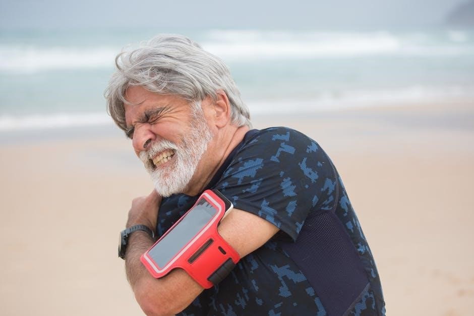
Understanding the plexus’s vulnerability during athletic activities, like football, is crucial for prevention and prompt intervention. Recognizing early symptoms and initiating appropriate management can significantly impact long-term outcomes.
Types of Brachial Plexus Injuries (Neuropraxia, Axonotmesis, Neurotmesis)
Brachial plexus injuries are classified by severity: neuropraxia (mildest), axonotmesis (axon damage, potential recovery), and neurotmesis (complete nerve severance, often requiring surgery). Neuropraxia, common in athletes, often resolves with conservative management, allowing return to play once symptoms subside. However, recurrent neuropraxia warrants investigation for underlying cervical pathology.
Axonotmesis and neurotmesis require more intensive interventions. Accurate diagnosis dictates the rehabilitation approach, ranging from NMES and progressive strengthening to surgical repair and prolonged post-operative therapy.
Initial Assessment & Diagnostic Tools
Comprehensive evaluation includes range of motion, strength testing, FG/SD curves, EMG/NCS, and imaging (radiographs/MRI) to pinpoint injury location and severity.
Clinical Evaluation: Range of Motion, Strength Testing
A meticulous clinical evaluation forms the cornerstone of brachial plexus injury assessment. Range of motion (ROM) testing assesses joint mobility, identifying limitations impacting function. Strength testing, utilizing manual muscle testing (MMT) scales, quantifies muscle weakness across affected myotomes.
Documenting active and passive ROM is crucial, noting any pain or resistance. Strength is graded on a 0-5 scale, revealing the extent of nerve involvement. Regular, repeated assessments are vital to track progress and adjust rehabilitation protocols accordingly. This data informs treatment planning and guides exercise selection, ensuring a tailored approach to recovery.
Functional Grading (FG) Test & Sensory Discrimination (SD) Curve
The Functional Grading (FG) test objectively assesses residual muscle function, crucial for guiding rehabilitation intensity. It categorizes muscle strength into grades, informing neuromuscular electrical stimulation (NMES) parameter adjustments. Simultaneously, the Sensory Discrimination (SD) curve evaluates sensory recovery, pinpointing areas of diminished sensation.
Regular FG and SD assessments—at intervals—allow therapists to modify exercises based on strength changes and sensory improvements. This dynamic approach optimizes treatment, promoting nerve regeneration and functional gains. Interpreting these tests enhances inter-professional communication, ensuring a coordinated recovery plan.
Electromyography (EMG) and Nerve Conduction Studies (NCS)
Electromyography (EMG) and Nerve Conduction Studies (NCS) are vital diagnostic tools for brachial plexus injuries, objectively assessing nerve and muscle function. NCS measure the speed of electrical signals, identifying nerve damage locations and severity. EMG evaluates muscle electrical activity, revealing denervation or reinnervation patterns.
These studies help differentiate injury types (neuropraxia, axonotmesis, neurotmesis), guiding treatment decisions. For chronic symptoms or recurrent episodes, EMG/NCS can rule out cervical pathology. Results inform prognosis and monitor rehabilitation progress, ensuring appropriate exercise prescription;
Imaging: Radiographs & MRI for Cervical Pathology
Radiographs and Magnetic Resonance Imaging (MRI) are crucial for identifying underlying cervical spine issues contributing to brachial plexus injuries. Radiographs detect fractures, dislocations, or bony abnormalities. MRI provides detailed soft tissue visualization, revealing nerve root compression, hematomas, or tumors.
Imaging is particularly important in athletes with chronic symptoms or repeated neuropraxia episodes, as highlighted in case reports. Ruling out cervical pathology guides appropriate management, preventing further nerve damage and optimizing rehabilitation protocols. Accurate diagnosis ensures targeted exercise prescription.
Phase 1: Acute Management & Early Rehabilitation (0-6 Weeks)
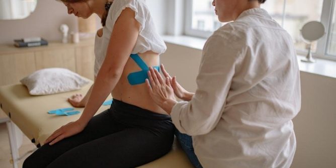
Initial focus involves pain control and gentle range of motion exercises, alongside neuromuscular electrical stimulation (NMES) with carefully adjusted settings.
Pain Management Strategies
Effective pain management is paramount during the acute phase of brachial plexus injury. Initial strategies often involve a combination of pharmacological interventions, such as analgesics, and non-pharmacological approaches. Gentle range of motion exercises, performed within pain-free limits, can help alleviate discomfort and prevent secondary complications like joint stiffness.
Furthermore, neuromuscular electrical stimulation (NMES), initiated with conservative settings, may contribute to pain modulation by stimulating nerve fibers and reducing muscle spasms. Careful monitoring of the patient’s pain levels and adjustments to the treatment plan are crucial. A multidisciplinary approach, involving physicians, physical therapists, and potentially pain specialists, ensures comprehensive pain control.
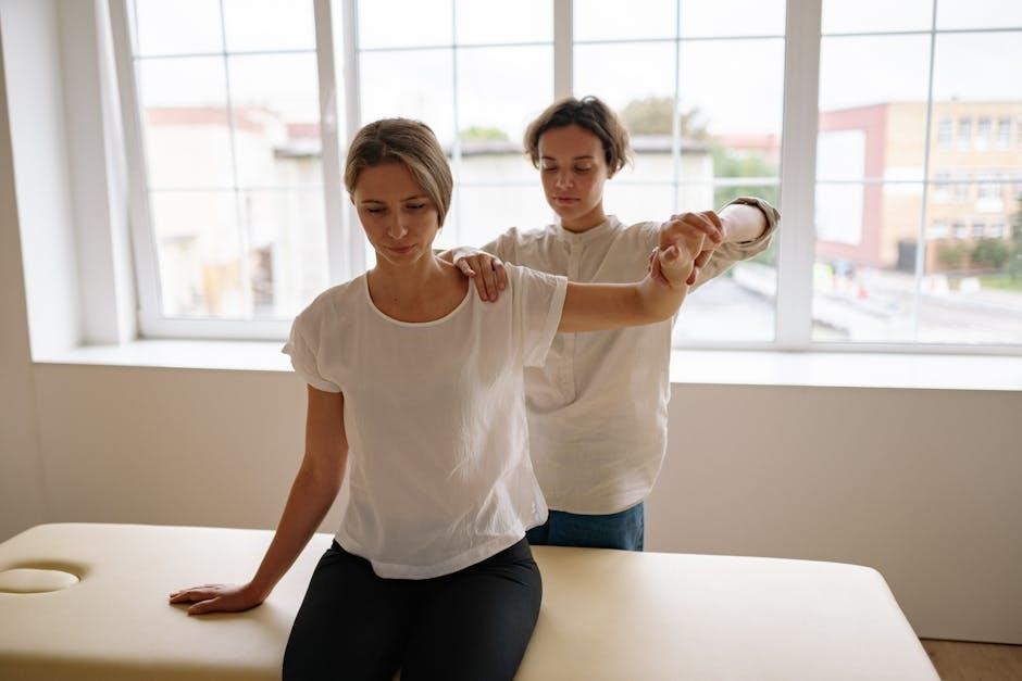
Gentle Range of Motion Exercises
Early implementation of gentle range of motion (ROM) exercises is vital to prevent secondary complications like contractures and stiffness following brachial plexus injury. These exercises should be performed passively or with minimal active assistance, staying within the patient’s pain tolerance. Focus initially on maintaining mobility in the shoulder, elbow, wrist, and hand.
Careful attention must be paid to avoid overstressing the injured nerves or muscles. The goal is to promote circulation, reduce swelling, and preserve joint integrity. Regular, controlled movements, guided by a physical therapist, are essential for optimizing recovery and preparing for more advanced rehabilitation phases.
Neuromuscular Electrical Stimulation (NMES) ─ Initial Settings
Neuromuscular Electrical Stimulation (NMES) plays a crucial role in early brachial plexus rehabilitation, aiming to re-educate weakened muscles and prevent atrophy. Initial settings should be conservative, utilizing symmetrical biphasic waveforms with a frequency between 20-50 Hz and pulse duration of 100-200 microseconds.
Intensity is gradually increased to elicit a visible muscle contraction without causing pain. Frequent monitoring of the Functional Grading (FG) test and Sensory Discrimination (SD) curve is essential to adjust NMES parameters, ensuring optimal stimulation and maximizing therapeutic benefits throughout the recovery process.
Phase 2: Intermediate Rehabilitation (6-12 Weeks)
Progressive strengthening, scapular stabilization, and active-assisted range of motion exercises are key during this phase, adapting to evolving muscle strength gains.
Progressive Strengthening Exercises
Initiating progressive strengthening requires careful monitoring of the Functional Grading (FG) test and Sensory Discrimination (SD) curve results. Exercises should begin with low resistance, gradually increasing as muscle strength improves. Resistance bands are excellent for providing adjustable resistance, allowing for controlled progression.
Focus on exercises targeting shoulder abduction, external rotation, elbow flexion and extension, wrist extension and flexion, and grip strength. Ensure proper form to avoid compensatory movements. Regular reassessment using FG/SD is crucial to modify exercises and prevent overexertion. The goal is to restore functional strength and endurance, preparing the patient for advanced rehabilitation phases.
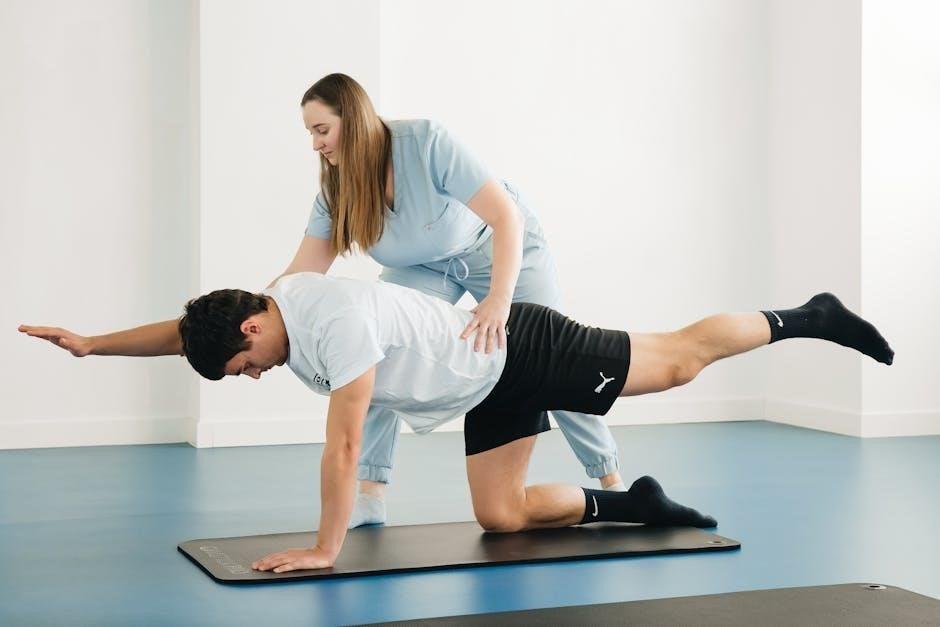
Scapular Stabilization Exercises
Robust scapular control is fundamental for optimal shoulder function following brachial plexus injury. Exercises should focus on strengthening the muscles responsible for scapular retraction, protraction, upward rotation, and downward rotation. These include the serratus anterior, trapezius, and rhomboids.
Begin with isometric exercises, progressing to dynamic movements like scapular squeezes, rows, and push-ups (modified as needed). Integrating these exercises with progressive strengthening enhances overall shoulder stability and facilitates functional movements. Consistent monitoring of patient progress, guided by FG/SD assessments, is vital for tailoring the exercise program.
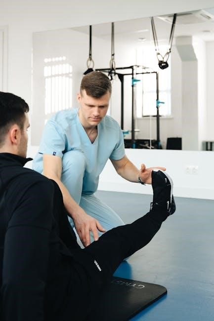
Active-Assisted Range of Motion (AAROM)
AAROM bridges the gap between passive range of motion and independent movement, crucial during intermediate brachial plexus rehabilitation. Utilizing the patient’s active effort combined with external assistance—from a therapist or assistive device—promotes neuromuscular re-education.
Focus on gentle, controlled movements, respecting pain boundaries. Exercises should target all shoulder planes, emphasizing scapulohumeral rhythm. Regularly reassess muscle strength changes using FG testing to modify exercise intensity and complexity, ensuring progressive gains in functional ability and preventing overexertion.
Phase 3: Advanced Rehabilitation & Functional Training (3+ Months)
Advanced training integrates resistance, proprioception, and task-specific drills to restore function, preparing patients for activities of daily living and potential sport return.
Advanced Strengthening with Resistance Bands & Weights
Progressive resistance training is paramount in Phase 3, utilizing bands and weights to rebuild strength lost due to nerve injury. Begin with light resistance, focusing on controlled movements and proper form to avoid re-injury. Exercises should target all affected muscle groups – shoulder abduction/adduction, external/internal rotation, elbow flexion/extension, and wrist movements.
Gradually increase resistance as strength improves, monitored by functional grading and patient feedback. Incorporate multi-planar exercises to mimic real-life activities. Careful attention to scapular stabilization is crucial throughout, ensuring optimal biomechanics. Remember, individualized programs are key, adapting to each patient’s specific deficits and progress.
Proprioceptive Training & Coordination Exercises
Restoring proprioception – the body’s awareness of its position in space – is vital for functional recovery after brachial plexus injury. Exercises should challenge balance and coordination, utilizing unstable surfaces like wobble boards or foam pads. Incorporate reaching tasks, weight shifting, and dynamic movements to improve neuromuscular control.
Coordination drills, such as throwing and catching, or simulated ADLs, enhance motor planning and execution. Focus on smooth, controlled movements, minimizing compensatory strategies. Progress from simple to complex exercises, continually challenging the patient’s ability to sense and react to changes in position.
Task-Specific Training for Activities of Daily Living (ADL)
Transitioning rehabilitation to real-world tasks is crucial for regaining independence. Task-specific training involves practicing ADLs – like dressing, eating, and grooming – with modifications as needed. Begin with simplified versions, gradually increasing complexity as strength and coordination improve.
Simulate work or sport-specific activities, focusing on movements essential for the patient’s lifestyle. Adaptive equipment may be incorporated to facilitate participation. This approach promotes neuroplasticity and ensures skills transfer effectively, maximizing functional outcomes and patient satisfaction.
Adaptive Equipment & Orthotic Aids
Selecting appropriate aids is crucial for rehabilitation; orthoses provide support and enhance function, enabling patients to participate more fully in daily activities.
Selecting Appropriate Adaptive Equipment
Careful consideration is paramount when choosing adaptive equipment for individuals with brachial plexus injuries. The goal is to maximize independence and participation in activities of daily living (ADL). Equipment selection must be individualized, based on the specific nerve root involvement, functional limitations, and patient goals.
Assistive devices can range from simple modifications, like built-up handles on utensils, to more complex solutions such as specialized reaching aids or environmental control systems. Orthotic aids, including splints and braces, play a vital role in supporting weakened muscles and improving joint stability. A thorough assessment by an occupational therapist is essential to determine the most appropriate equipment to enhance function and quality of life.
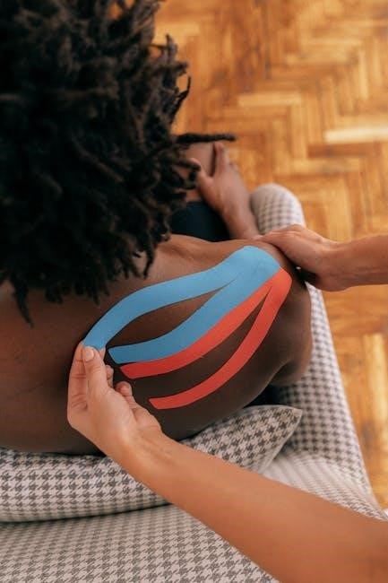
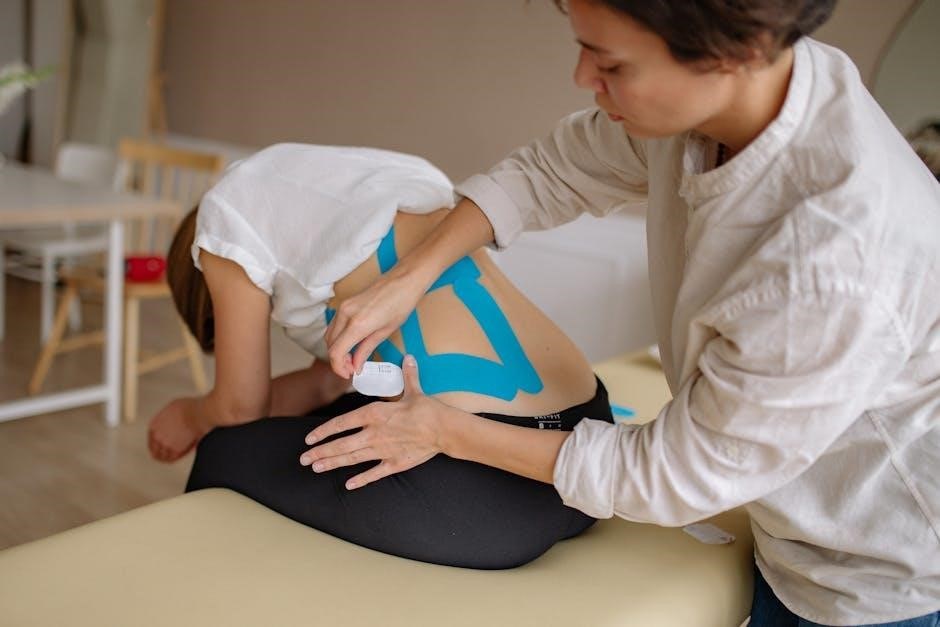
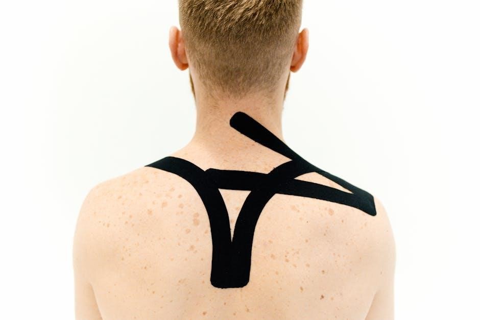
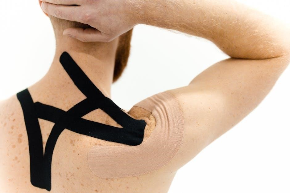
Use of Orthoses for Support & Function
Orthoses are integral to brachial plexus injury rehabilitation, providing crucial support and facilitating functional recovery. They address muscle weakness, joint instability, and limited range of motion. Dynamic orthoses, allowing movement while providing support, are often preferred during the intermediate phases of rehabilitation.
Static orthoses offer stability, particularly when neuromuscular control is severely compromised. Custom-fabricated orthoses ensure optimal fit and effectiveness. The use of orthoses should be integrated with a progressive exercise program to promote muscle strengthening and functional gains, ultimately maximizing the patient’s ability to perform daily tasks;
Return to Sport Considerations
Athletes returning after brachial plexus neuropraxia require a gradual protocol, symptom monitoring, and consideration of previous episodes before resuming full activity.
Gradual Return to Activity Protocol
A phased return to sport is paramount following brachial plexus injury. Initial stages focus on pain management and regaining full, pain-free range of motion. Progressive strengthening, incorporating scapular stabilization exercises, follows, building a foundation for functional movements. Athletes then advance to proprioceptive training and coordination drills.
Crucially, return to play is contingent on a normal clinical examination and complete symptom resolution. Monitoring for recurrence is vital, as repeated neuropraxia episodes can occur if athletes return too soon. Radiographs and MRI may be needed for chronic symptoms, alongside EMG assessments.
Neuropraxia & Return to Play Guidelines
Return to play after brachial plexus neuropraxia generally occurs once symptoms resolve and a clinical examination is normal. This can sometimes even happen within the same game, though consensus remains elusive. However, athletes often delay reporting symptoms, leading to repeated neuropraxia before seeking medical attention.
Careful monitoring is essential. If symptoms persist for weeks, radiographs or MRI should investigate cervical pathology, and electromyography may be considered. A gradual return to activity protocol, prioritizing full recovery, minimizes the risk of recurrence and ensures long-term athlete well-being.
Monitoring for Recurrence of Symptoms
Vigilant monitoring post-rehabilitation is crucial, as athletes may attempt premature return to play, potentially triggering repeated neuropraxia episodes. Regular clinical evaluations, including range of motion and strength testing, are essential to detect subtle changes.
Promptly address any symptom resurgence—pain, weakness, or sensory alterations—with immediate activity modification. Continued intercommunication between medical professionals ensures a coordinated response. Proactive management minimizes chronic complications and optimizes long-term functional outcomes, safeguarding the athlete’s health and performance.
Long-Term Management & Prevention
Sustained exercise programs and consistent interprofessional communication are vital for addressing chronic symptoms and preventing future brachial plexus injury occurrences.
Ongoing Exercise Program
A consistent, tailored exercise program is paramount for long-term brachial plexus injury management. This should incorporate scapular stabilization, progressive strengthening with resistance bands and weights, and proprioceptive drills to restore coordination.
Regular neuromuscular electrical stimulation (NMES), guided by Functional Grading (FG) and Sensory Discrimination (SD) curve assessments, helps maintain muscle activation. Adapting exercises based on strength gains is crucial.
The program must address potential chronic symptoms and complications, emphasizing adherence and proactive adjustments. Intercommunication between surgeons, physiatrists, and physiotherapists ensures optimal, individualized care and sustained functional improvement.
Importance of Intercommunication Between Medical Professionals
Effective brachial plexus injury recovery hinges on seamless communication between surgeons, physiatrists, and physiotherapists. Collaborative assessment, utilizing FG tests and SD curves, guides rehabilitation protocol modifications based on evolving muscle strength.
Regular dialogue ensures NMES settings are optimized, and exercise progressions are appropriately timed. This interdisciplinary approach facilitates earlier recovery than anticipated, addressing individual patient needs.
Shared expertise minimizes complications and promotes a holistic, patient-centered strategy, ultimately maximizing functional outcomes and long-term well-being.
Addressing Chronic Symptoms & Potential Complications
Persistent brachial plexus symptoms necessitate investigating cervical pathology via radiographs or MRI, especially in athletes with recurrent neuropraxia episodes. Electromyography aids in diagnosing prolonged symptoms exceeding several weeks.
Managing chronic pain and functional limitations requires ongoing exercise programs tailored to individual needs. Athletes may attempt premature return to play, exacerbating injuries; therefore, careful monitoring is crucial.
Proactive management minimizes long-term disability, focusing on adaptive equipment, orthotic support, and addressing any emerging complications to optimize quality of life.
Case Study Analysis: Rehabilitation Successes
College and high school football players demonstrate successful recovery through tailored rehabilitation protocols, emphasizing early intervention and interdisciplinary communication for optimal outcomes.
Review of Case Reports – College Football Player
A case report detailed a college football player with complete C5 and C6 nerve root avulsion. Rehabilitation focused on meticulous clinical evaluation, utilizing the Functional Grading (FG) test and Sensory Discrimination (SD) curve to guide neuromuscular electrical stimulation (NMES) adjustments;
Exercise modifications were implemented as muscle strength improved, demonstrating the importance of a dynamic protocol. Successful recovery hinged on consistent intercommunication between surgeons, physiatrists, and physiotherapists. This collaborative approach facilitated earlier-than-expected recovery, highlighting the benefits of a structured, adaptable rehabilitation program tailored to the athlete’s specific needs and progress.
Review of Case Reports – High School Football Player
A high school football player’s case illustrated the challenges of chronic brachial plexus neuropraxia and cervical extension loss. The athlete initially downplayed symptoms, attempting to return to play prematurely, resulting in recurrent neuropraxia episodes. Diagnostic imaging, including radiographs and MRI, ruled out cervical pathology, while electromyography aided in assessing symptom duration.
Rehabilitation emphasized addressing chronic symptoms and preventing further injury. Return-to-play guidelines were strictly followed, contingent upon complete symptom resolution and a normal clinical examination, acknowledging the lack of universal consensus on immediate return.
Resources & Further Information
Explore the RGUHS Journal of Physiotherapy, PMC, and Acibadem Health Point for comprehensive insights into brachial plexus injury management and rehabilitation protocols.
Relevant Journals (RGUHS Journal of Physiotherapy, PMC)
The RGUHS Journal of Physiotherapy provides valuable clinical perspectives on brachial plexus injury rehabilitation, highlighting the importance of regular Functional Grading (FG) and Sensory Discrimination (SD) curve assessments. These journals emphasize adapting neuromuscular electrical stimulation (NMES) settings based on these evaluations and modifying exercises as muscle strength improves.
PMC (PubMed Central) offers access to case reports, such as those detailing chronic neuropraxia in athletes, informing return-to-play decisions and the need for thorough cervical pathology investigation via imaging when symptoms persist. Both resources promote interprofessional communication for enhanced patient outcomes.
Acibadem Health Point Resources
Acibadem Health Group emphasizes a crucial aspect of brachial plexus rehabilitation: the strategic selection of adaptive equipment and orthotic aids. These resources underscore the necessity of tailoring assistive devices to individual patient needs, maximizing functional recovery and supporting optimal movement patterns.
Their protocols align with a comprehensive approach, integrating these aids into a broader rehabilitation plan that includes progressive exercises and task-specific training. Acibadem’s focus on personalized care ensures patients regain independence and improve their quality of life post-injury.